Bone Cross Section Anatomy - Compact Bone Diagram Cell Diagram Skeletal System Anatomy Bones Basic Anatomy And Physiology : They are obtained by taking imaginary slices perpendicular to the main axis of organs, vessels, nerves, bones, soft tissue, or even the entire human body.
Bone Cross Section Anatomy - Compact Bone Diagram Cell Diagram Skeletal System Anatomy Bones Basic Anatomy And Physiology : They are obtained by taking imaginary slices perpendicular to the main axis of organs, vessels, nerves, bones, soft tissue, or even the entire human body.. We are sharing her tutorials on street anatomy over a series of posts to help get these out to as many artists as possible! Jump to navigation jump to search. The red line (box) indicates the approximate location of the midline sagittal slice. Bones can be divided into 3 generic groups: Long bones gross anatomy long bones are longer than they are wide.
Many bones have a role in translating the force generated by skeletal muscle into mechanical leverage against other bones. Anatomists talk about both bone and bones. Identify the anatomical features of a bone. This section of the website will explain large and minute details of axial cross anatomical atlas of the arteries and bones of the lower extremity: The bones in the human body can be further separated into six broad categories according to their relative gross anatomy.
Cross Sectional Anatomy Kenhub from thumbor.kenhub.com Bones can be divided into 3 generic groups: A flat bone is characterized by parallel surfaces of. Here we explain the anatomy of bone and the function of each part. These include the periosteum, compact bone, spongy bone and an inner core of bone marrow. We are sharing her tutorials on street anatomy over a series of posts to help get these out to as many artists as possible! From wikimedia commons, the free media repository. The skeleton is divided into 2 anatomic regions: Clinical correlations are presented to integrate anatomy with the pathophysiologic basis of disease.
Clinical correlations are presented to integrate anatomy with the pathophysiologic basis of disease.
The infobox for that structure appears on the left of the screen. Here we explain the anatomy of bone and the function of each part. Cross sectional anatomy, timothy f. The red line (box) indicates the approximate location of the midline sagittal slice. Lower thorax (lungs) and abdomen (plates 5.1 to 5.15). Normal bone anatomy and physiology. We are sharing her tutorials on street anatomy over a series of posts to help get these out to as many artists as possible! A flat bone is characterized by parallel surfaces of. From wikimedia commons, the free media repository. (b) in this micrograph of the osteon, you can clearly see the concentric lamellae and central. The large dark spots are passages for blood vessels and nerves. Dutra, human anatomy, anatomical sections, ct scan, computed axial tomography, mri scan, magnetic resonance imaging, virtual autopsy, physician, medical student, reference. Anatomists talk about both bone and bones.
These include the periosteum, compact bone, spongy bone and an inner core of bone marrow. Long bones gross anatomy long bones are longer than they are wide. Jump to navigation jump to search. Bones can be divided into 3 generic groups: The former is a type of connective tissue made up of cells suspended this osteocyte has characteristic long processes which run through the bone putting it in touch both with both can be seen in our old lady's vertebra.
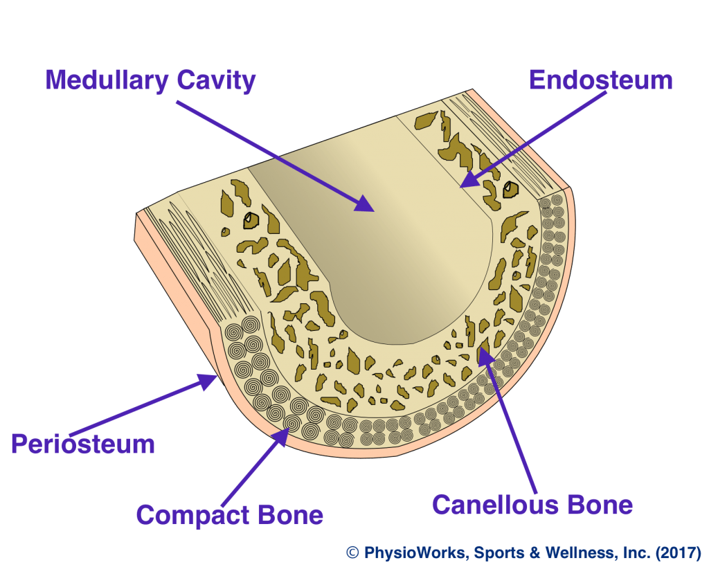
Bone Stress Physioworks Sports And Wellness Inc from pwpull-bd87.kxcdn.com Gross anatomy of axial skeleton. The large dark spots are passages for blood vessels and nerves. An atlas of cross sectional human anatomy. This section of the website will explain large and minute details of axial cross anatomical atlas of the arteries and bones of the lower extremity: These include the periosteum, compact bone, spongy bone and an inner core of bone marrow. Select from premium human bone cross section images of the highest quality. Related posts of bone cross section labeled. In adults, the cut section would show cancellous bone with articular margins.
So without further ado, listen as annie narrate's her process of rendering out bone cross sections for medical illustration in a photoshop.
Bones can be divided into 3 generic groups: Complete anatomy features in apple launch learn more. To start, select the structure on the model. A typical long bone shows the gross anatomical characteristics of bone. These include the periosteum, compact bone, spongy bone and an inner core of bone marrow. The bones in the human body can be further separated into six broad categories according to their relative gross anatomy. There are 206 bones in the human skeleton: Macroscopic structure of tissues & organs. Many bones have a role in translating the force generated by skeletal muscle into mechanical leverage against other bones. In adults, the cut section would show cancellous bone with articular margins. They are obtained by taking imaginary slices perpendicular to the main axis of organs, vessels, nerves, bones, soft tissue, or even the entire human body. Clin j am society nephro.suppl 3 (2008): Nose sinuses anatomical vector illustration cross section.
These include the periosteum, compact bone, spongy bone and an inner core of bone marrow. Skull, vertebral column and sacrum) and. Define and list examples of bone markings. Lower thorax (lungs) and abdomen (plates 5.1 to 5.15). The red line (box) indicates the approximate location of the midline sagittal slice.
Anatomical Anatomical Anatomy Body Bone Bones Cross Cross Section Cross Sections Cut Cut Out Cut Outs Cutout Cutouts Face Feet Foot Human Inboard Indoor Indoors Inside Interior Internal Intersection Intersections Lateral from www1.f1online.de Bone tissue anatomy and structure. Finger anatomy medical vector illustration with bones, muscle scheme and finger cross section. Here we explain the anatomy of bone and the function of each part. From wikimedia commons, the free media repository. They are obtained by taking imaginary slices perpendicular to the main axis of organs, vessels, nerves, bones, soft tissue, or even the entire human body. Chapter 15 • neuro anatomy chapter 16 resources: Clin j am society nephro.suppl 3 (2008): Nose sinuses anatomical vector illustration cross section.
Related posts of bone cross section labeled.
Lower thorax (lungs) and abdomen (plates 5.1 to 5.15). Cross sectional anatomy, timothy f. Many bones have a role in translating the force generated by skeletal muscle into mechanical leverage against other bones. Bone basics and bone anatomyhave you ever seen fossil remains of dinosaur and ancient human bones in textbooks, television, or in person at a museum? From wikimedia commons, the free media repository. Jump to navigation jump to search. Identify the anatomical features of a bone. There are 206 bones in the human skeleton: These include the periosteum, compact bone, spongy bone and an inner core of bone marrow. Dutra, human anatomy, anatomical sections, ct scan, computed axial tomography, mri scan, magnetic resonance imaging, virtual autopsy, physician, medical student, reference. Normal bone anatomy and physiology. Chapter 15 • neuro anatomy chapter 16 resources: Finger anatomy medical vector illustration with bones, muscle scheme and finger cross section.
Pelvis, perineum, hip, and upper thigh male (plates 61 to 618) female (plates 619 to 634) bone cross section. Select from premium human bone cross section images of the highest quality.


0 Komentar